Revolutionizing Pathology with Infrared-Optical Hybrid Microscopy
In modern pathology, traditional microscopy faces a significant limitation: it requires specialized staining techniques to visualize tissue structures and composition. While infrared (IR) imaging offers a potential solution by creating virtual tissue stains, its compatibility with standard optical microscopes is hindered by the absorption of IR light by many optical components. This limitation makes IR imaging incompatible with existing research and clinical settings.
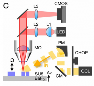
To address this challenge, Martin Schnell and his team developed the Infrared-Optical Hybrid (IR-OH) method, which combines optical imaging with infrared absorption analysis to determine tissue composition and structure. Their groundbreaking work demonstrates that this hybrid approach outperforms state-of-the-art IR microscopy in several key areas. Notably, it enables high-resolution imaging of unstained tissues at the microscopic level while also allowing for computationally generated stains based on these images.
The implementation of IR-OH microscopy relies heavily on advanced digital imaging technology. Adimec’s gentificTM High Full Well camera (Q-2HFW) was used to capture tissue images in this research, showcasing its role in enabling the hybrid approach. According to Martin Schnell, the Q-2HFW’s exceptional full-well capacity and signal-to-noise ratio were critical for handling the demanding data acquisition requirements of IR-OH imaging.
Technical Specifications
The Adimec Q-2HFW-CXP camera features:
- 14400×1440 resolution at up to 550 fps frame rates based on 12-micron pixels
- Global shutter CMOS image sensor optimized for maximum full-well performance
- Full well capacity exceeding over 2 million electrons per pixel
- Unique signal-to-noise ratio (SNR) performance of approximately 63dB
- Resolution: 1440×1440 with CoaXPress interface
- Frame rate capability up to 550 fps
- Global shutter CMOS sensor optimized for maximum full-well performance
The paper describing this innovative approach was published in Proceedings of the National Academy of Sciences. The camera’s unique specifications make it particularly well-suited for IR-OH applications:
- Full-well capacity: Over 2 million electrons per pixel
- Resolution: 1440×1440 at up to 550 fps
- Unique SNR performance delivering approximately 63dB
Advantages of the Hybrid Approach
The innovative method has significant implications for both research and clinical pathology. By overcoming IR absorption limitations, this technology could transform how pathologists analyze tissues by enabling:
- Simultaneous analysis of tissue morphology and biochemical composition
- Real-time image processing capabilities
- Compatibility with existing optical microscopy workflows
This breakthrough technology offers a potentially more efficient alternative to traditional staining methods while providing deeper insights into tissue composition. The IR-OH method’s ability to capture bright scenes without saturation, combined with its compatibility with standard laboratory equipment, addresses previous technical limitations that had stalled development since 2010.
Implementation Challenges
The research team discovered in the Q-2HFW camera due to:
- Deep-pixel sensors preventing sensor over-saturation even under intense light conditions
- Ability to stream data directly from camera to computer for real-time processing
This capability was essential for handling massive datasets—up to 1000 TB generated during a 7-day continuous operation period.
Future Implications
The IR-OH method offers substantial advantages over traditional techniques:
- Cheaper alternative to conventional staining methods
- Shorter processing times and reduced labor requirements
The hybrid approach can transform pathology practices by enabling comprehensive tissue analysis without reliance on potentially toxic chemical stains
This innovative technology is poised to reshape pathological research and clinical diagnosis, providing a powerful new tool for understanding tissue composition and structure. For more details about this revolutionary imaging solution, visit the original paper here: https://www.pnas.org/content/117/7/3388
Last Updated: 2025-09-04 18:26:34