Development of High-Performance CMOS Camera for Real-Time Cancer Imaging
Adimec (Netherlands), in collaboration with five partners within the CAReIOCA consortium, is advancing a high-resolution, high-speed imaging medical device designed for non-invasive optical biopsy assessments in cancer diagnostics. Adimec contributes its expertise through a specialized high-performance CMOS CoaXPress camera.
The initiative began early 2013, and initial results were unveiled in June 2014. Additional updates are featured in the third newsletter. Notable achievements from late 2014 include:
- Integration of a CMOSIS sensor into an ADIMEC camera.
- Completion by Leiden University Medical Center (LUMC) and Institut Gustave Roussy (IGR) of clinical atlases comparing FFOCT and histological images for breast and head & neck cancer biopsies.
- Development by LLTech of two FFOCT imaging device prototypes, with a preliminary endoscope version set to launch in December 2014.
The overarching aim of the CAReIOCA project is to supply pathologists and surgeons with real-time, non-invasive optical cellular-level imaging within the human body. The core technology relies on Full Field Optical Coherence Tomography (FFOCT), capable of capturing volumetric images of semi-transparent tissues at micron resolution in 3D. This innovation seeks to aid cancer diagnosis across various organs—skin, breast, prostate, brain—and support biopsy quality assurance.
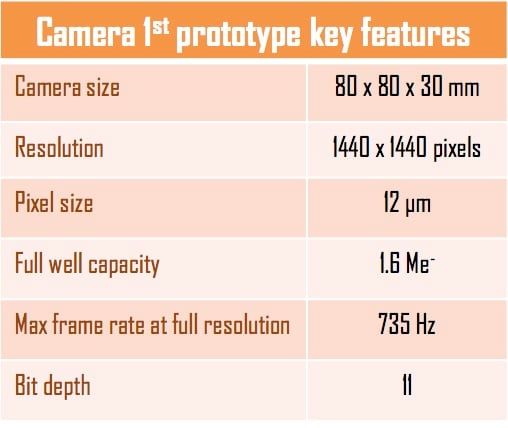
High-Speed CMOS CoaXPress Camera Development
ADIMEC constructed the first camera prototype and shipped it to LLTech mid-July. The image sensor underwent characterization by CMOSIS before being integrated into Adimec’s camera platform, ensuring high-speed precision imaging and seamless interfacing for easy incorporation into LLTech’s FFOCT devices. This CoaXPress interface facilitates efficient handling of high-bandwidth data streams.
Further prototyping persists at both CMOSIS and ADIMEC. LLTech has embedded the initial prototype into their LightCT scanner for preliminary testing, highlighting benefits like superior full-well capacity leading to minimized shot noise. For instance, FFOCT camera prototypes exhibit a significantly enhanced signal-to-noise ratio (SNR), potentially tripling gains with further tuning.
Clinical Atlas Completion
LUMC in Leiden and IGR in Villejuif spearheaded the creation of clinical atlases—comprehensive databases pairing FFOCT images with histology for breast cancer research at LUMC and head & neck cancer studies at IGR. These resources, displayed alongside annotated slides, are pivotal for diagnostic training and refinement.
FFOCT Device Prototypes
LLTech finalized designs for two high-resolution microscopes derived from the LightCT scanner by incorporating new cameras and refining sample holders/travel mechanics/software. The contact endoscope prototype uses a novel tandem interferometry optical system enabling probe interchangeability in a compact unit, prioritizing image quality and size.
This project was funded under FP7-ICT grant agreement number 318729 from the European Union’s Seventh Framework Program for Information Technology. For further details on CAReIOCA updates, visit Click here.
Last Updated: 2025-09-04 20:18:22