Revolutionizing Digital Pathology with High-Resolution Imaging Technology
A significant demographic shift toward aging populations in many countries has led to an increase in cancer and other diseases. This trend results in a higher volume of patients and samples, while the number of skilled pathologists does not grow at the same pace. Additionally, there is a strong push—particularly in oncology—to shorten diagnosis times for better treatment outcomes.
Pathology laboratories must enhance output and efficiency without compromising quality. Digital pathology systems, including automated digital microscopes and slide scanners, enable the digitization of images once viewed manually. These technologies offer numerous benefits: reducing diagnostic time, allowing remote expert consultations (telepathology) without physical slide shipment, enabling efficient data storage, and more.
Adimec’s 12-megapixel cameras (Q-12A65 and Q-12A180) provide high-resolution imaging with fast frame speeds to meet the demands of these systems. The exceptional image quality ensures reliable input for further analysis. Features like region-of-interest (ROI) functionality allow users to adjust sharpness settings quickly and efficiently before capturing full-resolution images.
Furthermore, forward-facing optical coherence tomography (FFOCT) complements traditional histology by being applicable during surgical procedures. Below is an example comparing FFOCT and histology (provided by LLTech).
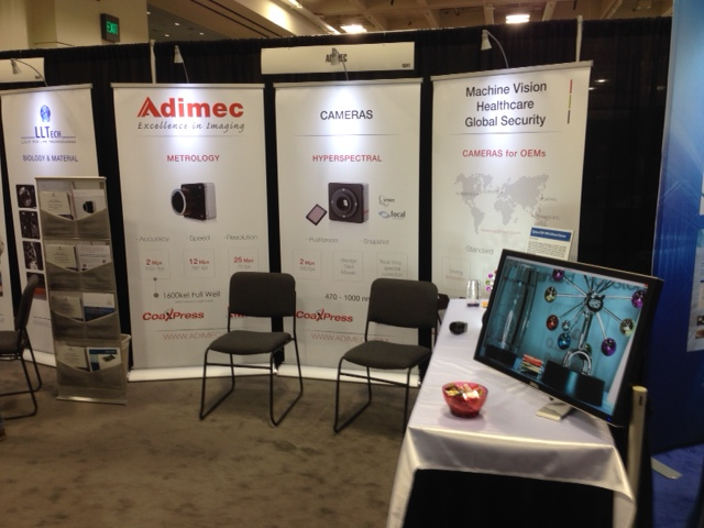
For more information on Adimec’s extreme full-well cameras or other models, visit us at booth #4541 at Photonics West this week.
Last Updated: 2025-09-04 20:22:12