Revolutionizing Bone Marrow Analysis with AI-Powered Digital Pathology
For serious blood diseases like leukemia, multiple myeloma, and lymphoma, differential counting of blood cells in bone marrow smears remains the diagnostic gold standard. Currently, these morphological assessments are performed manually by pathologists and highly-trained lab personnel, requiring significant concentration and precision from technicians. Human factors such as stress, fatigue, distraction, and training level can introduce errors into test interpretations—scientists refer to this as “inter-operator variation.” To enhance accuracy and efficiency in diagnostic hematology, aetherAI has developed Microscope x Hema—a complete digital pathology system that uses The Imaging Source’s USB 3.0 color microscopy cameras to create digital images processed with deep learning techniques.
Cell Classification via Deep Learning
To properly train its CNNs, aetherAI partnered with the National Taiwan University Hospital to develop the world’s first differential-counting AI model for bone marrow smears. Trained on a comprehensive dataset of 500,000 annotated samples, Microscope x Hema’s embedded solution includes AI-powered microscope control software, an AI model for differential counting, and dedicated hardware supporting AI inferencing.
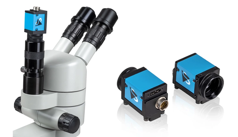
Standard optical microscopes often produce images with complex backgrounds that hinder efficient cell analysis. The system uses the 20 MP DFK 33UX183 camera’s high-sensitivity CMOS sensor to deliver low-noise images (high signal-to-noise ratio). Its image pre-processing reduces residual noise, enhancing edges and contours while highlighting details and reducing blur.
Microscope x Hema’s algorithms extract features from the images and set parameters such as shape, contour, irregular fragments, color, and texture. The workflow is complete once the system classifies and counts all cells in the sample.
Differentiation and Categorization of Nucleated Bone Marrow Cells
Using aetherAI’s Microscope x Hema analysis platform—which processes images captured by DFK 33UX183 cameras—healthcare professionals can differentiate and categorize nucleated bone marrow cells with greater ease. The software displays pre-processed images of bone marrow cells along with reports on the percentage and number of cell categories.
AetherAI’s Vision for Digital Pathology
By easing the burden on healthcare professionals, aetherAI aims to improve medical diagnostics through “solutions for digital pathology and AI-powered diagnostic support.” Company founder Dr. Joe Yeh stated, “The AI revolution will realize the ultimate value of digital medical images and bring healthcare to the next level.”
Last Updated: 2025-09-05 00:18:59