Comparison with a Leica Add-on Camera
Although comparing two different cameras directly can be challenging due to the numerous variables that influence results, it is still meaningful to compare our test subject with the Leica MC 170 HD for reference purposes. We began by photographing neurons from specimens shown in Figures 8, 9 and 11 using the original 40x Plan-corrected objective on the Leica microscope DMLB equipped with its built-in camera. The camera was mounted manufacturer-specified onto the C-Mount head of the trinocular photo tube. Manual adjustment was required because the Leica camera lacked One-Push autofocus.
For comparison, we used the same Plan-corrected 40x objective with a camera/eyepiece combination from The Imaging Source to image neurons from the same specimens (using selective One Push autofocus).
The Leica camera provided an image resolution of 2592 x 1807 pixels. We selected the highest possible resolution setting (4128 x 3096 pixels) for the The Imaging Source camera.
Neurons appeared significantly smaller when captured by the later system with its 10x eyepiece compared to those captured by the Leica camera and its special projection lens. To facilitate comparison, we extracted a section from the larger image (4128 x 3096 pixels) that exactly matched the original dimensions of the Leica camera’s output (2592 x 1807 pixels). Now both images showed neurons at nearly identical magnification.
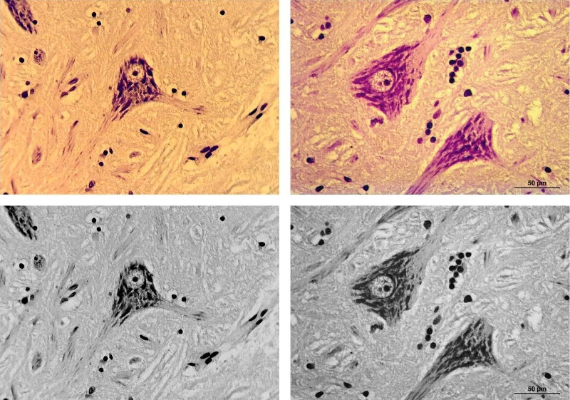
Figure 17 presents these comparison images alongside optimized black-and-white versions. This demonstrates that for routine tasks like documenting bright-field images, cameras from The Imaging Source can compete effectively with leading manufacturer solutions in terms of image quality.
Figure 17
Neurons captured via two different systems: left panel shows the test camera system combined with Meiji eyepiece; right panel uses Leica MC 170 HD. Images are displayed above (color) and below (black-and-white).
Last Updated: 2025-09-05 01:00:30