Illumination Types and Filters
In addition to bright-field images, techniques such as polarized light, darkfield, and phase contrast imaging can be flawlessly captured using the camera. Even very narrowly filtered monochromatic light (red, green, and blue) posed no issues for the autofocus system—neither in terms of exposure nor precision. Objects illuminated with a single wavelength produced clear black-and-white images.
Figures 13 through 16 showcase various applications taken under different lighting conditions, filter types, and microscope setups:
-
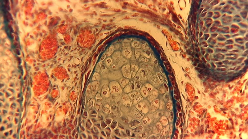
Figure 13: Bright-field image captured with the Meiji eyepiece on a Carl Zeiss Jena large-format tube paired with a Leitz plan-corrected NPL 40/0.65 objective lens, showing rat embryo tissue (cartilagenous vertebral column). Despite lacking an eyepiece compensator, results remain usable.
-
Figure 14: Darkfield imaging using the Meiji eyepiece and Carl Zeiss Jena large-format tube with a Leitz plan-corrected scanning objective. The sample is xylit crystal without cover slip, illuminated by unfiltered halogen light (top), ‘minusviolet’ filter (middle), or monochromatic green 540 nm filter (bottom). Autofocus is activated via One-Push.
-
Figure 15: Polarized light imaging with the Meiji eyepiece and Carl Zeiss Jena large-format tube equipped with a Leitz plan-corrected scanning objective. Xylit crystal without cover slip, using a lambda compensator for polarization effects. Autofocus enabled by One-Push.
-
Figure 16: Polarization imaging similar to Figure 15 but without the compensator lens, again on Carl Zeiss Jena equipment with Leitz objectives and automated autofocus via One-Push.
Last Updated: 2025-09-05 01:01:05