JAI Launches New Camera Models and Software Integrations for Microscopy Systems
JAI has introduced a series of new camera models and software integrations that enhance color imaging capabilities for microscopy-based systems. The announcement includes six new variations within the Apex Series of high-performance 3-CMOS prism color cameras, along with full integration into two popular microscopy software solutions: Image-Pro® from Media Cybernetics and µManager open-source software.
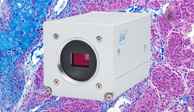
New Camera Models
Three models are based on the AP-3200T-USB, a 3.2-megapixel camera operating at 38.3 frames per second (fps). The other three models derive from the AP-1600T-USB, a 1.6-megapixel device offering higher frame rates but lower resolution. All cameras feature USB3 Vision interfaces, known for their bandwidth and plug-and-play compatibility.
The new variants include standard green housings alongside white options designed for clinical or laboratory environments where white is often preferred. Some models lack standard IR-cut filters to enhance red sensitivity, useful in life sciences applications requiring detailed differentiation of stains or tissues. These cameras are also beneficial for food and industrial analyses involving simultaneous visible and near-infrared (NIR) imaging.
Software Integration
JAI has partnered with Media Cybernetics to integrate the Apex Series cameras with Image-Pro software, enabling seamless image transfer and control. For users preferring open-source solutions, the µManager platform now supports JAI cameras through custom device adapters, available without licensing fees from micro-manager.org.
Applications
The new models’ C-mount compatibility allows integration with most digital microscopes. Their 3-CMOS prism technology delivers superior color differentiation compared to Bayer sensors in brightfield and fluorescence imaging. Higher frame rates support real-time applications like live cell imaging and time-lapse microscopy.
These cameras excel in diverse fields, including pathology for tissue analysis and stain evaluation; materials science for defect classification in coatings or composites; wafer inspection for semiconductor defects; and more. Specific use cases include:
- Live Cell Imaging: The 38 fps or 79 fps frame rates support time-lapse microscopy.
- Pathology: High color fidelity aids tissue slice analysis, stain evaluation, and cell classification.
- Fluorescence Microscopy: Enhanced red sensitivity supports disease diagnosis and neuron signaling studies.
- Materials Science: Accurate color imaging benefits cosmetic and composite material analysis.
- Wafer Inspection: High resolution ensures precise defect detection in semiconductor manufacturing.
Last Updated: 2025-09-05 02:48:56